Cone Beam CT Imaging Studies and Services delivered securely to your desktop…
At Orbit, we specialize in delivering high-quality diagnostic imaging and reporting services for dental and maxillofacial needs. Our expert team leverages advanced imaging technologies to handle the entire process—from image acquisition to preparation and detailed reporting—allowing you and your staff to concentrate fully on patient care. We provide secure, efficient access to images and reports, empowering you to deliver the highest standards of care to your patients. Trust Orbit to be your partner in precision imaging and exceptional patient support.
3D Imaging Studies
-
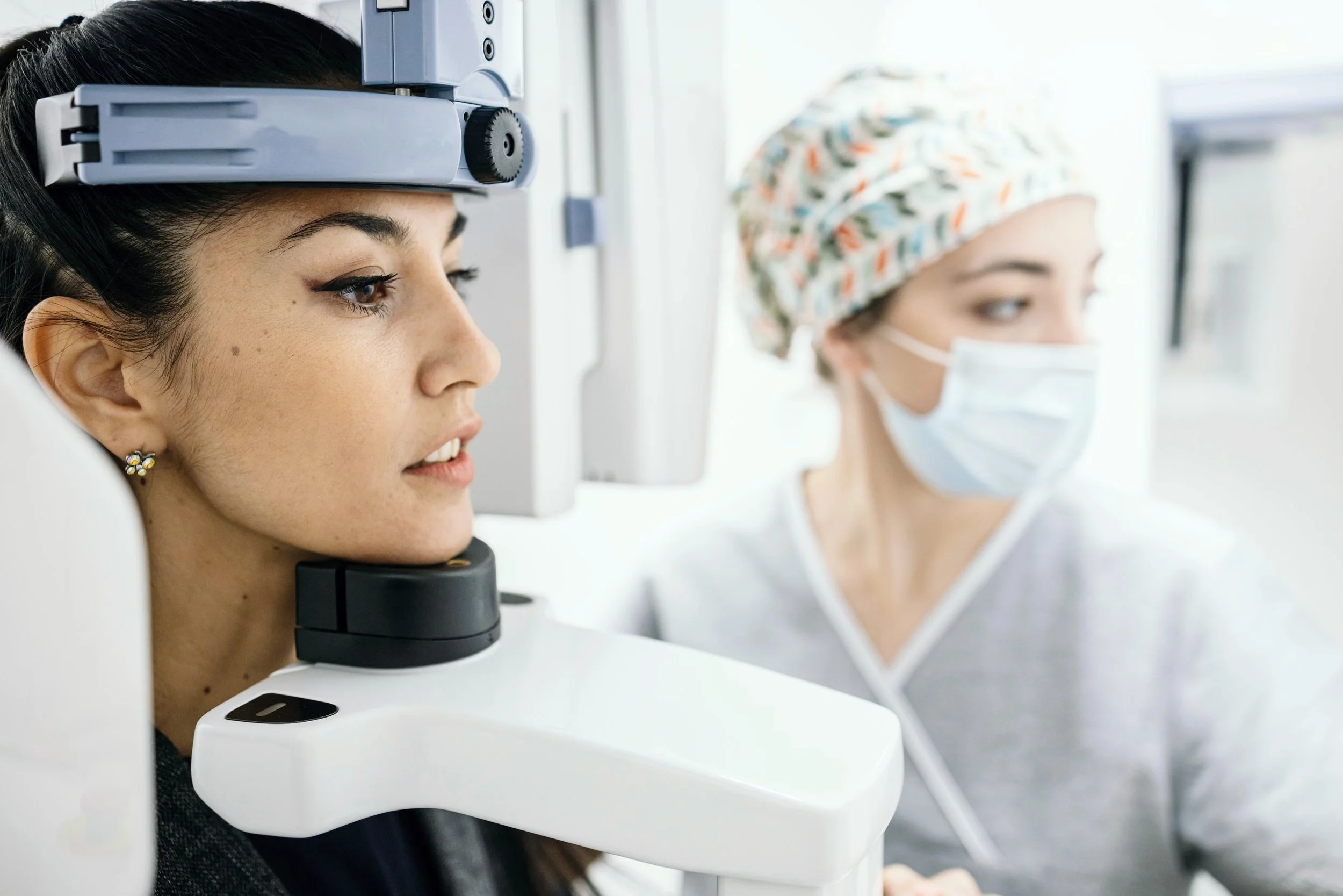
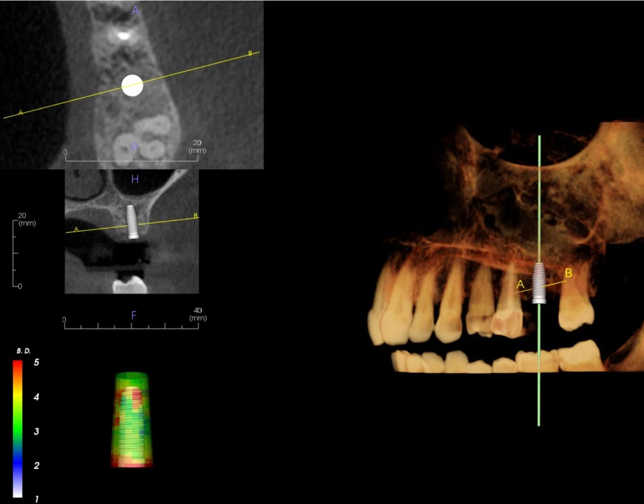
We deploy a variety of scanners allowing for a variety of fields of view from focus field, to full head.
What is a Cone-Beam Computed Tomography or CBCT? Is it a CT scan?
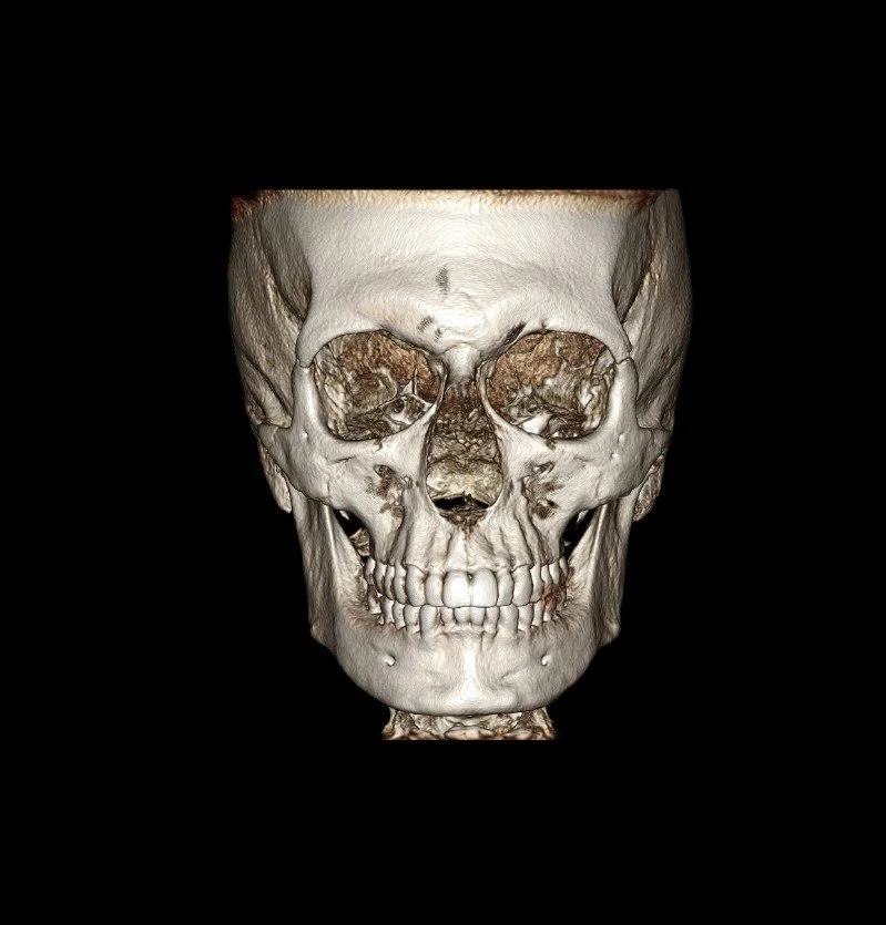
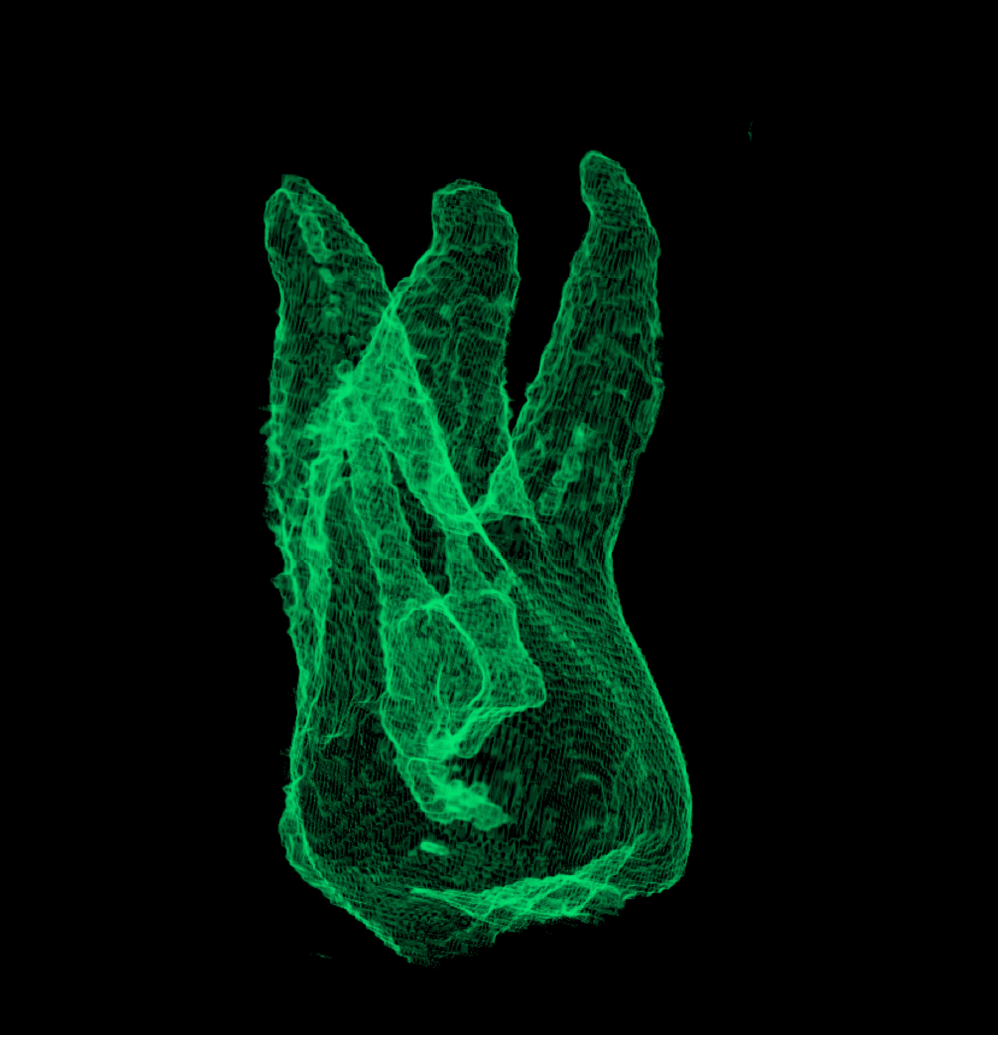
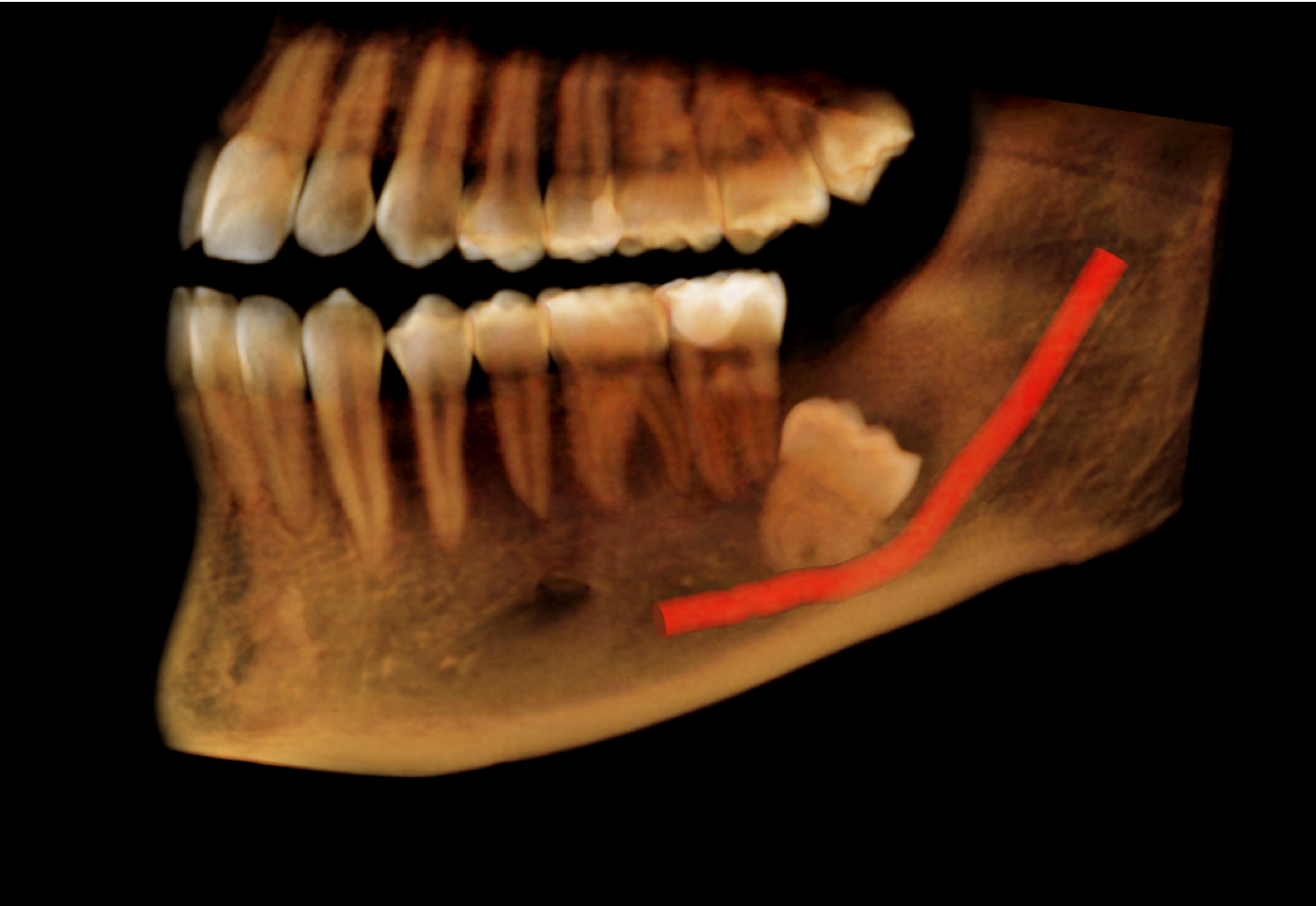
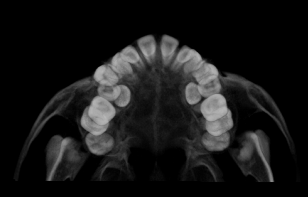
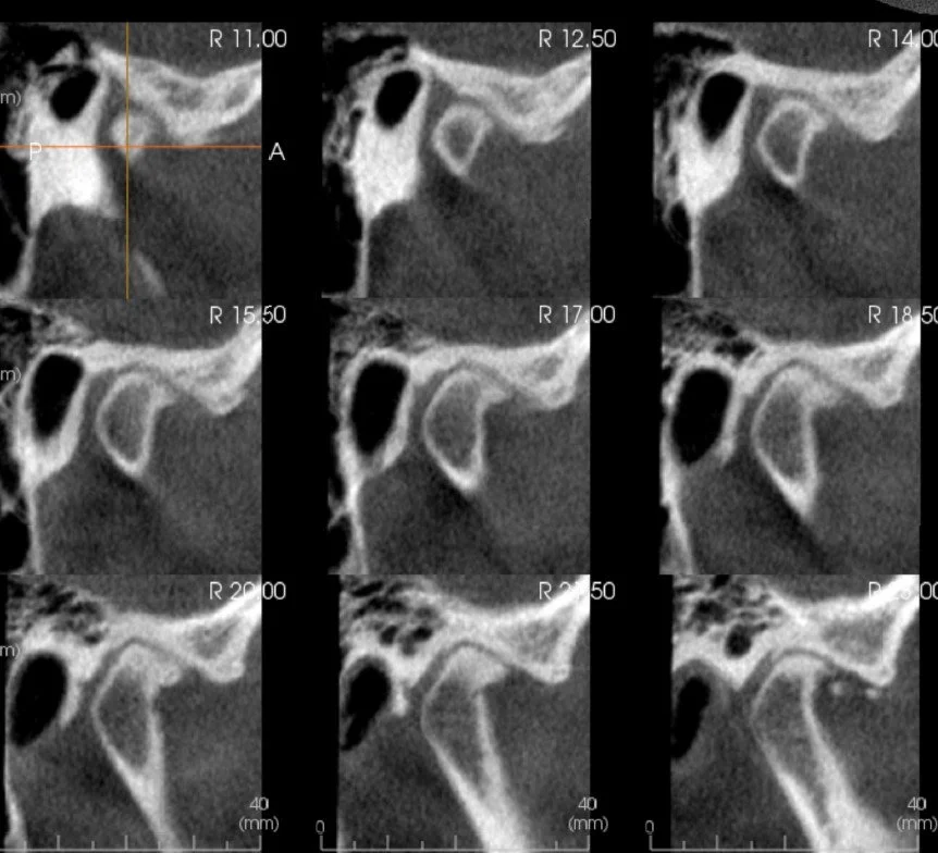
Cone-beam Computed Tomography, or CBCT for short, is a type of low-dose CT scan that allows us to visualize your teeth, jaws, TM joints and airways accurately in 3-dimensions. This advanced imaging technology has markedly improved our ability to diagnose and treat several dental and jaw problems. Dental CBCT examinations expose you to considerably less radiation than the CT scans made in hospital settings.
-
Intraoral scanners (IOS) are used to create detailed digital impressions of a patient's teeth and oral structures. IOS offer many advantages over putty based impressions. Applications include:
1. Invisalign / Orthodontic Planning & Treatment: Creating precise models for designing braces, aligners, and other orthodontic appliances.
2. Implant Planning: Assisting in the planning and placement of dental implants.
3. Monitoring Treatment: Tracking changes over time and evaluating the progress of treatments.
Intraoral scanners improve accuracy, efficiency, and patient comfort, making them a valuable tool in modern dental practice.
Dental Imaging Services
2D Imaging Studies
-
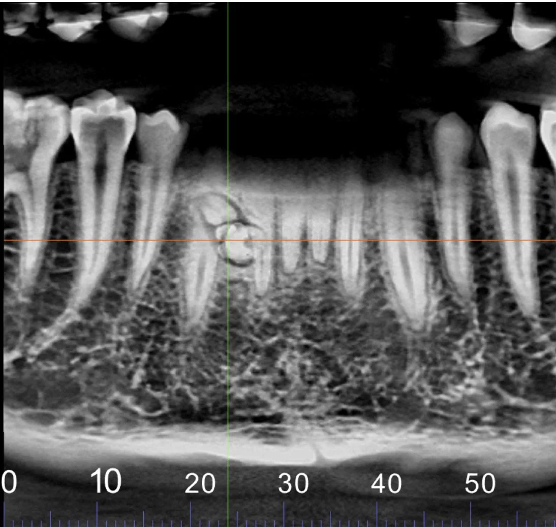
Panoramic images are digital 2D dental x-ray examinations that captures the teeth, jaws and TMJs in a single image. The panoramic is an overview image that is used to visualize erupted and unerupted teeth, evaluate root alignment, TMJ’s and the sinuses.
-
Cephalometric images are a critical diagnostic tool in orthodontics and maxillofacial treatment. The cephalometric image, provides a detailed side or front to back view of the head's skeletal, dental and facial structures. It is most commonly used in treatment planning, identification of airway issues and analysis of growth and development.
-
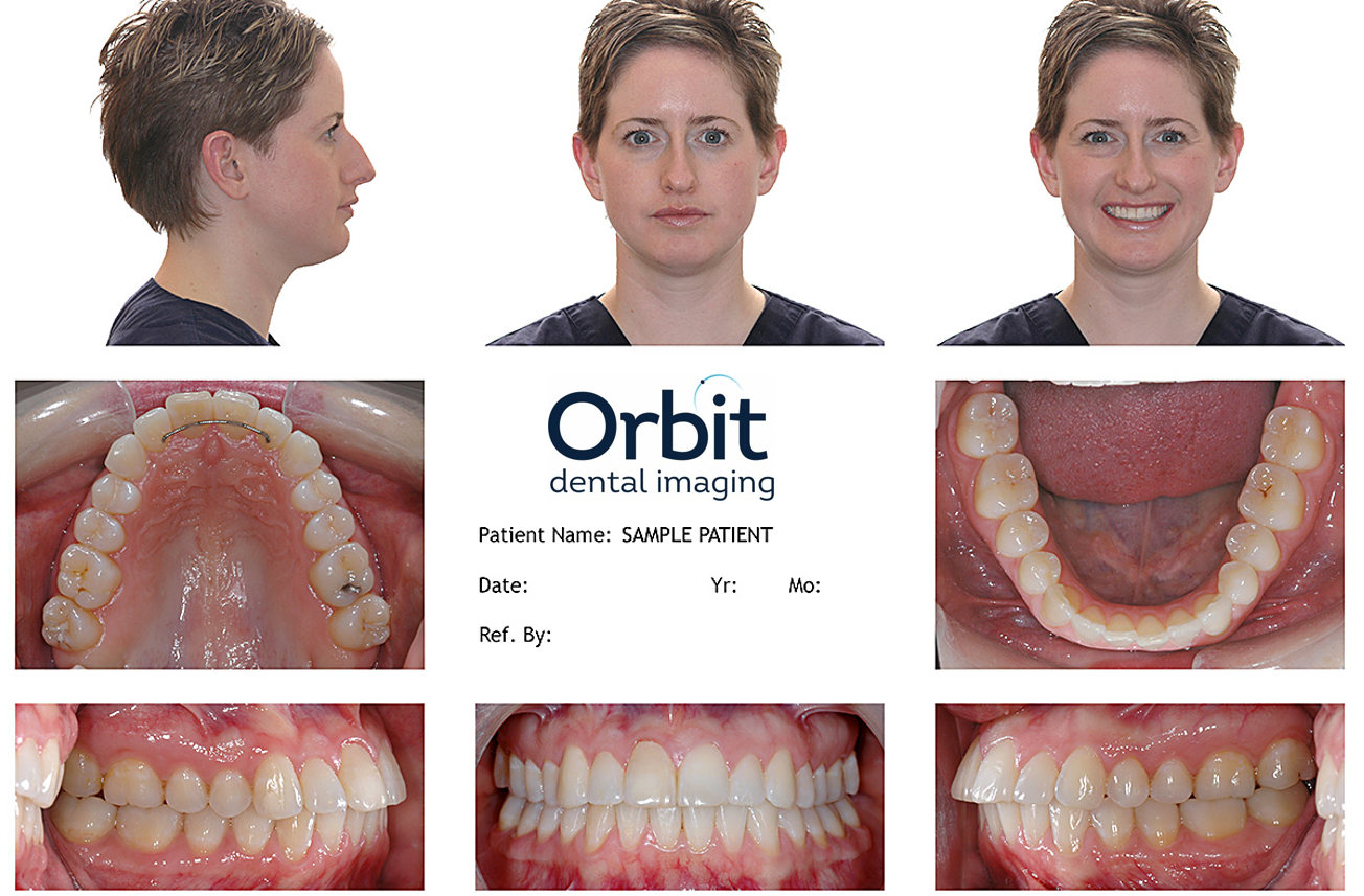
Clinical photographs are used for documenting the condition of a patient's teeth, face and oral structures for diagnostic, treatment planning, and educational purposes.
Imaging Reports
-
An Oral and Maxillofacial Radiology (OMFR) report is crucial in the diagnosis and treatment planning for conditions affecting the head, neck, face, jaws, and oral structures. The value of such a report includes:
1. Accurate Diagnosis: It provides detailed imaging that helps in accurately diagnosing complex dental and maxillofacial conditions, including tumors, cysts, fractures, and developmental abnormalities.
2. Treatment Planning: The report guides dentists, oral surgeons, and other healthcare providers in creating precise and effective treatment plans, ensuring that all aspects of a patient's condition are addressed.
3. Early Detection: Radiology reports can detect issues that might not be visible during a clinical examination, allowing for early intervention, which can improve patient outcomes.
4. Legal and Medical Documentation: The report serves as a vital part of the patient's medical records, which can be used for legal purposes or for future reference in ongoing treatment.
5. Communication: It facilitates clear communication among multidisciplinary teams, ensuring that all practitioners involved in a patient’s care are on the same page regarding the diagnosis and treatment plan.
Overall, an OMFR report is a key tool in ensuring comprehensive patient care in dental and maxillofacial practice.
-
An image portfolio report translates the 3D volume into clear and organized image sets enabling patient communication in a way they will understand. Images are correctly oriented and have true size and scaling for optimum planning.
Image portfolio reports are fully customizable to give you the information you need to prepare and communicate your treatment plan.
-
Tracing and analysis of a lateral cephalometric radiograph allows clinician to assess craniofacial relationships, dental alignment, and skeletal growth patterns. It helps orthodontists and surgeons diagnose malocclusions, plan treatments, and monitor progress by providing detailed measurements of the jaws, teeth, and facial bones. This analysis supports accurate treatment planning for braces, jaw surgeries, and other corrective procedures.
Implant Planning & Guides
-
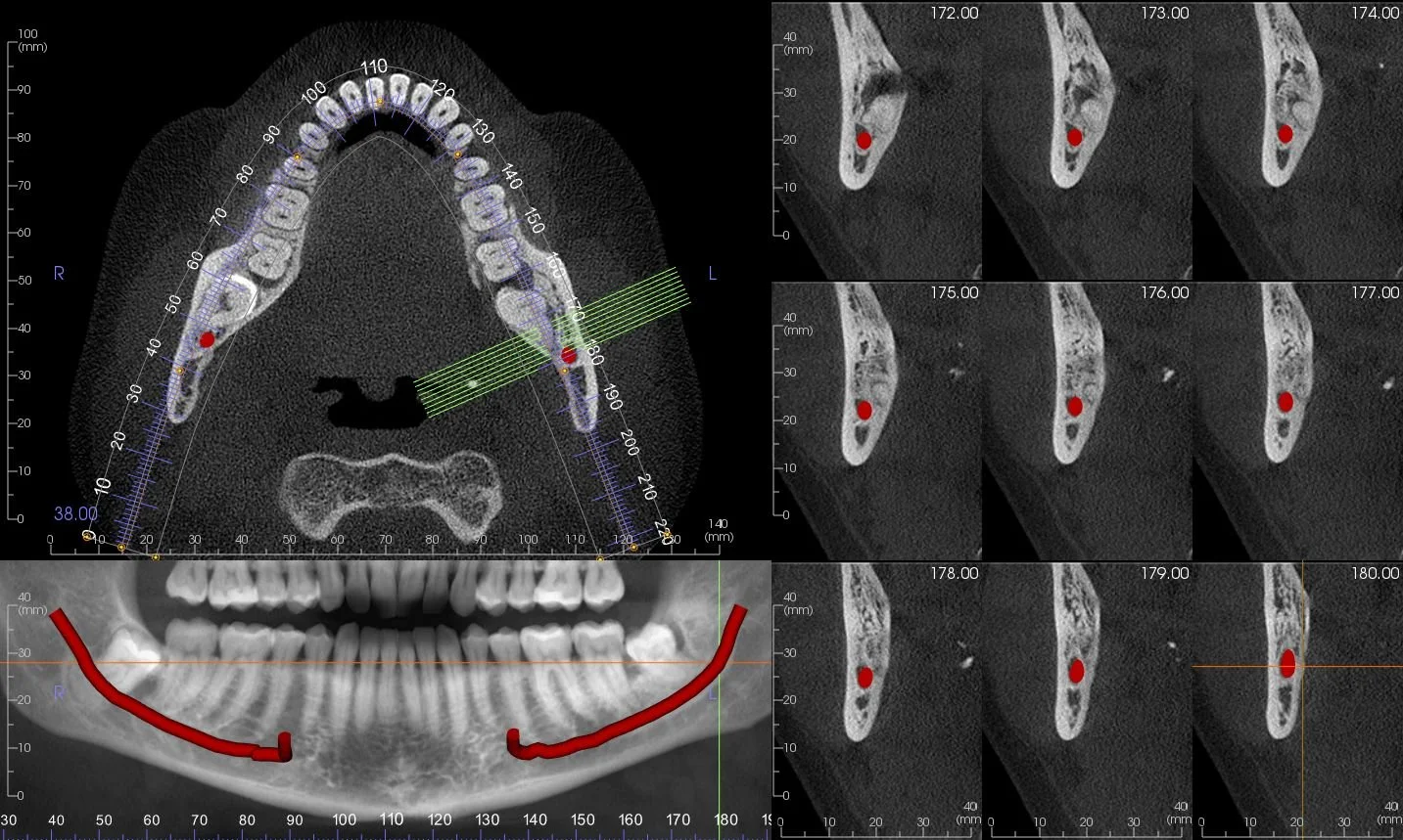
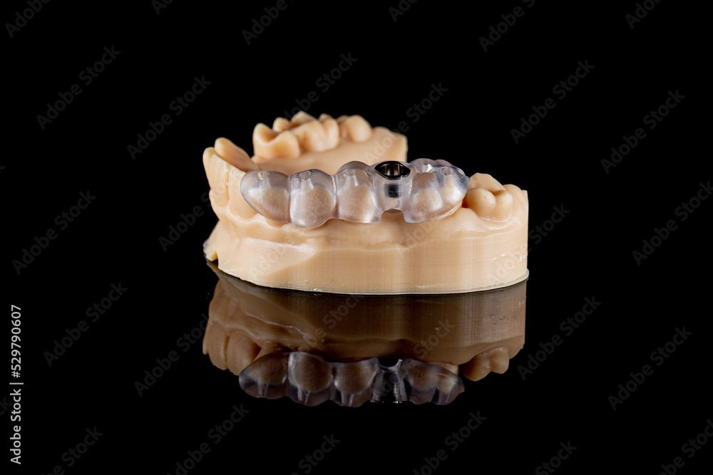
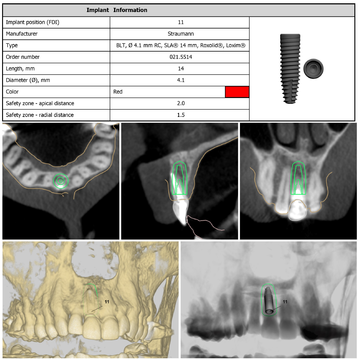
Our surgical guides are custom, patient specific devices designed and created from 3D imaging data to assist implant placement during dental implant surgery. The purpose is to guide the precise placement of dental implants within the patient’s jawbone with optimal accuracy thereby minimizing the risk of any errors or misplacement.
-
Orbit's Implant Plans are created in 3 Shape Implant Studio planning environment. by an experienced dental professional in collaboration with the implant dentist or specialist. The plans are prosthetically driven and include an interactive planning session with the referring clinician. The resulting plan can be used to create a surgical guide by Orbit or or by a preferred dental laboratory.
Sample Report Gallery

